From Massive to Miniature: How Ultrasound Technology Has Changed Towards Portability & User-Friendly
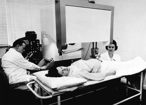
Ultrasound machines have traveled a long path after its invention. The introductory ultrasound machine was a 2-dimensional scanner introduced in 1958 by Tom Brown and Ian Donald. They called it the Diasonograph, but it was well-known in more informal and unkind circles as the Dinosaurograph,

Fig. 1 Ian Donald operating the Mark 3 Diasonograph
This ascendant of the current day ultrasound device was 8 ft tall and occupied close to a third of the space available in an average scanning room. The ultrasound images were still and were formed by rocking the transducer slowly over the body part that needs to be examined. This was no small effort as the gantry that housed the probe was very large as well, and much physical exertion had to be utilized to alter the scanning plane. The image wholly lacked any greyscale, but it would still look eventually on a cathode ray tube. The Diasonograph’s first contribution to medicine and ultrasound technology is that it glint the ultrasound race.
By the early 1970s real-time machines had been manufactured. Companies such as Aloka and Kretztechnic then quickly changeover from these mechanical sectors real-time scanners to the multi-element linear array and phased array scanners as soon as the mid-70s. The color Doppler imaging option made its debut in 1985, at around the same time when Japanese researcher Kazunon Baba began experimenting with 3D imaging. 3D/4D ultrasound came into the market by the mid-1990s with the 530D Voluson. By the year 2000 one could find high-resolution harmonic imaging, 3D/4D capabilities, and power Doppler facilities in most ultrasound machines.
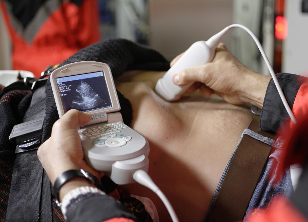
In comparison to the competition forever more progressive characteristic, there was another race of size of it. The big ultrasound machines of old were rapidly replaced by smaller consoles. The first portable ultrasound unit came into the market in the United States as early as 1975 from ADR (which was finally purchased by Philips). It consists of 3 linear probes and weighed only 25 pounds. Soon battery powered portable ultrasound machines were fabricated as well, and now ultrasound machines that can able to fit into one’s pocket are commercially available. The first such latest piece of a medical device was the Siemens Acuson P10 Hand-held Ultrasound Machine. It weighs barely 1.6 lbs (.725kg).

Fig. 2 The Handheld Ultrasound Machine
Companies such as GE, Philips, and Abbot are all competing to be the manufacturers of the smallest machines with the top-grade resolution. The gain of portable ultrasound machines are so on and varied, and engineers at such companies are ambitious at work to convert them ever more portable. The quality of images even on portable machines is phenomenal, and they have many of the characteristics that full ultrasound consoles do, yet engineers are working to bridge the small gap between consoles and portable machines as well.
Ultrasound machines have developed into sleek tools that are used to provide detailed images of what lies underneath the skin. It now seems infeasible to imagine obstetrics without ultrasound technology. Life-threatening heart conditions are diagnosed and then treated by applying ultrasound machines in echocardiography every hour. Even potential future dangers are being foretold by using ultrasound to screen for blood vessel disease.Ultrasound probes have gone from a gargantuan rarity to a tiny necessity, and the future day of ultrasound technology looks bright.
For more details visit: http://u.to/bORDEQ

Fig. 1 Ian Donald operating the Mark 3 Diasonograph
This ascendant of the current day ultrasound device was 8 ft tall and occupied close to a third of the space available in an average scanning room. The ultrasound images were still and were formed by rocking the transducer slowly over the body part that needs to be examined. This was no small effort as the gantry that housed the probe was very large as well, and much physical exertion had to be utilized to alter the scanning plane. The image wholly lacked any greyscale, but it would still look eventually on a cathode ray tube. The Diasonograph’s first contribution to medicine and ultrasound technology is that it glint the ultrasound race.
By the early 1970s real-time machines had been manufactured. Companies such as Aloka and Kretztechnic then quickly changeover from these mechanical sectors real-time scanners to the multi-element linear array and phased array scanners as soon as the mid-70s. The color Doppler imaging option made its debut in 1985, at around the same time when Japanese researcher Kazunon Baba began experimenting with 3D imaging. 3D/4D ultrasound came into the market by the mid-1990s with the 530D Voluson. By the year 2000 one could find high-resolution harmonic imaging, 3D/4D capabilities, and power Doppler facilities in most ultrasound machines.
In comparison to the competition forever more progressive characteristic, there was another race of size of it. The big ultrasound machines of old were rapidly replaced by smaller consoles. The first portable ultrasound unit came into the market in the United States as early as 1975 from ADR (which was finally purchased by Philips). It consists of 3 linear probes and weighed only 25 pounds. Soon battery powered portable ultrasound machines were fabricated as well, and now ultrasound machines that can able to fit into one’s pocket are commercially available. The first such latest piece of a medical device was the Siemens Acuson P10 Hand-held Ultrasound Machine. It weighs barely 1.6 lbs (.725kg).

Fig. 2 The Handheld Ultrasound Machine
Companies such as GE, Philips, and Abbot are all competing to be the manufacturers of the smallest machines with the top-grade resolution. The gain of portable ultrasound machines are so on and varied, and engineers at such companies are ambitious at work to convert them ever more portable. The quality of images even on portable machines is phenomenal, and they have many of the characteristics that full ultrasound consoles do, yet engineers are working to bridge the small gap between consoles and portable machines as well.
Ultrasound machines have developed into sleek tools that are used to provide detailed images of what lies underneath the skin. It now seems infeasible to imagine obstetrics without ultrasound technology. Life-threatening heart conditions are diagnosed and then treated by applying ultrasound machines in echocardiography every hour. Even potential future dangers are being foretold by using ultrasound to screen for blood vessel disease.Ultrasound probes have gone from a gargantuan rarity to a tiny necessity, and the future day of ultrasound technology looks bright.
For more details visit: http://u.to/bORDEQ
Comments
Post a Comment